Pavilion Publishing and Media Ltd
Blue Sky Offices Shoreham, 25 Cecil Pashley Way, Shoreham-by-Sea, West Sussex, BN43 5FF, UNITED KINGDOM
Tel: +44 (0)1273 434 943
Email: [email protected]


Although skin problems form 10% of the GP workload, their diagnosis remains challenging. The consultation can be more difficult if it is a pregnancy specific dermatoses, often managed by the midwife or secondary care, rather than the GP – and especially so if they are the first point of contact. Nevertheless, the GP has an important role to play, as some problems can significantly affect both maternal and fetal health and alter the birth plan.
The aim of this article is to aid the GP in recognising pregnancy specific dermatoses, knowing what treatment can be initiated in primary care, and who to refer to in secondary care.
Skin conditions in pregnancy may be pre-existing, which may or may not be exacerbated by pregnancy, physiological, or pregnancy specific dermatoses.1 The key symptom in pregnancy specific dermatoses is pruritis, and the skin conditions are classified into four conditions as polymorphic eruption of pregnancy (PEP), atopic eruption of pregnancy (AEP), pemphigoid gestationis (PG) and intrahepatic cholestasis of pregnancy (ICP).2 One differentiating feature is whether a rash is present.
PEP is the most common pregnancy specific dermatoses, occurring in up to one in 160 pregnancies.3 It typically affects first pregnancies in the final trimester, or in 15% of cases in the postpartum period. It is more common in multiple pregnancies and overweight mothers.4 No hormonal or immunological cause has been discovered. An inflammatory reaction secondary to abdominal distention is one proposed theory.5

Pruritis is a prominent symptom with erythematous, polymorphous urticarial papules, which coalesce into plaques, initially on the abdomen striae before migrating to the buttocks and thigh, with distinctive periumbilical sparing (see Figure 1). PEP rarely affects the hands, feet and skin above the breasts. Small vesicles may develop later in the disease and a targetoid rash may occur.5
Initial management should be started by the GP. Regular topical emollients, a moderate potent steroid, an oral antihistamine and antipruritic such as menthol can be used. First generation oral antihistamines such as chlorphenamine are preferred, aiding sleep due to the sedatory effect, with patients advised to keep well hydrated to avoid any anti-cholinergic side effects. Cetirizine and loratidine can be offered if the patient wants a non-sedating option after the first trimester. Crusting and scaling is seen in six weeks.5
If response is inadequate, short-term oral steroids may be used, at high dose and tapered down quickly.7 Prior to this, a discussion with an obstetrician is recommended, with a follow-up in an antenatal clinic.8 Narrow band ultraviolet light B (NB-UVB), albeit rarely used in PEP, has been shown to be effective, requiring dermatology assessment.9
No maternal complications have been linked to PEP, which seldom recurs in subsequent pregnancies, except multiple pregnancies. Lesions characteristically resolve within six weeks postpartum.1 Infants may acquire atopic dermatitis later in life.10
AEP has an incidence of one in 300 and is triggered by an immunological change.9 Over 70% of presentations occur before the third trimester and as a primary condition, rather than a flare-up of a pre-existing dermatitis.2
The key clinical finding is dry skin.1 This group can be further divided into an eczematous type, with extensive eczema in characteristic atopic sites, including the flexures, face, palm and soles.2 The second type is papular, seen on the abdomen and extensor surfaces of the arms and legs, with prurigo nodules on the shins and arm.11
Initial treatment is with regular topical emollients, oral antihistamines, and antipruritics12 can be used.13 A topical moderate potent steroid should be initiated, moving to potent topical steroids in poor control.
Parallel to PEP, if a topical steroid is unsuccessful, then oral prednisolone can be used and tapered when controlled, following obstetric discussion with regular follow up in an antenatal clinic. NB-UVB has been shown to decrease intensity by 30%, necessitating dermatology assessment.14 Topical benzoyl peroxide and erythromycin have also been successful therapies in case reports, but require secondary care input.15
Maternal risk is low, although there is the possibility of symptoms relapse in subsequent pregnancies. The fetus is unaffected, but may acquire atopy in infancy2 care.
PG is a bullous, autoimmune dermatosis affecting one in 50,000 pregnancies, usually presenting after the first trimester. It is related to HLA DR3 and DR4 and has been linked to molar pregnancies and choriocarcionoma.16
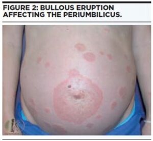
Initially, patients experience a marked burning sensation and pruritis, followed by periumbilical urticarial papules coalescing to plaques.1 Subsequently, a widespread bullous eruption occurs, involving the face and mucosa in 10% and 20% of cases respectively.17 The severity of PG is associated with early onset and blister development.18
Whist awaiting dermatology assessment, mild cases may be managed with topical steroids and oral antihistamines.19
To ensure correct diagnosis, histology and direct immunofluorescence is undertaken, and therefore a referral to a dermatologist is needed.20
The majority of cases require oral systemic steroids. If required, the dose of steroid should be reduced pre-term and increased postpartum. In refractory cases postpartum, dapsone, cyclophosphamide and methotrexate have been trialled with variable success and safety.16
An improvement is seen late in pregnancy, but in 75% of cases a recurrence is seen at delivery or postpartum resolving within months.21 Breastfeeding has been shown to reduce the duration of postpartum PG.22
A recurrence may also occur with reestablishment of the menstrual cycle or with oral contraceptive use.19 PG is seen in future pregnancies at earlier gestation with more severe features and longer duration postpartum.19 A link with other autoimmune diagnoses such as Graves disease and pernicious anaemia has been found,23 as well as conversion to bullous pemphigoid.24
PG is associated with premature labour and small for gestational age babies due to placental failure, so additional antenatal surveillance is recommended.25 Ten percent of babies develop transient blisters from passive maternal transfer of IgG antibodies. As with most other transient newborn diseases caused in this way, they resolve spontaneously within weeks.25
In multi-ethnic populations in England, ICP affects 0.7% of pregnancies,26 observed in the third trimester and linked to multiple gestation. The pathogenesis is multifactorial, a family history is noted in 50% of cases, and a link to oestrogen metabolites has been suggested.27
Patients develop abrupt onset pruritis, worst at night, on the palms of the hands and soles of the feet prior to becoming more generalised. Skin lesions appear secondary to patient scratching, typically as excoriations on the extensor aspects of the back, abdomens and limbs.25 ICP is the second most common cause of jaundice in pregnancy, observed in up to 15% of cases. Other symptoms include steatorrhea, dark urine, insomnia and lethargy.28
Laboratory investigations show elevated transaminases, possible deranged clotting function, with bile acids the most sensitive and specific marker for ICP,28 although 45% of ICP cases can have normal fasting bile acids.29 Furthermore, biochemical changes can occur weeks after symptom onset, so liver function tests (LFT) should be rechecked every 1-2 weeks in patients with continuing undiagnosed pruritis.30 There is an association with preterm labour and possibly with antepartum stillbirth.31
Blood tests can be checked in primary care, but it is advised that all patients with suspected ICP should be referred to obstetric care on the same day,32 with a liver screen and liver ultrasound completed.33 Regular emollients, menthol antipruritic and oral antihistamine may provide symptom relief.34
Ursodeoxycholic acid (UDCA) (500mg twice a day or 15mg/kg per day) is commonly prescribed, but a large trial35 showed that although it reduces itching, the effect is very small and for most patients not clinically worthwhile. The effect of UDCA on the fetus has been studied in a number of trials, but results are compatible with both important benefits and harms.36 Larger trials are ongoing. Cholestyramine, a bile acid chelator, can exacerbate vitamin K deficiency and has not been shown to improve fetal outcomes. If the36 prothrombin time is protracted then menadiol 5-10mg daily is indicated.33
Weekly maternal liver and clotting blood tests, weekly fetal cardiotocographic and regular growth scans from 30 weeks for fetal surveillance are warranted.37
The mainstay in resolution of symptoms of ICP is delivery of the baby. Induction is sometimes offered after 37 week of gestation, especially with severe biochemical changes, although in milder cases an active approach has limited evidence.33
Cholestasis can result in inadequate vitamin K and coagulopathy inducing a haemorrhage.19 A higher risk of gallstones has been observed in primigravida with ICP.28 ICP has a high recurrence rate – as high as 70% – in subsequent pregnancies.38 The fetal risks include premature delivery, meconium aspiration and possibly fetal demise.38 The cause may be secondary to noxious bile acids. As bile acid concentration increases, fetal risk increases.31
In primary care, LFT should be checked at 6 weeks and if an abnormality remains then a referral to gastroenterology be made. LFT should not be routinely offered in the first 10 days postpartum as they may still be deranged.33
Oestrogen based contraceptives can be started once biochemistry has normalised, but need review if cholestasis recurs.38
Mitesh Patel Academic Clinical Fellow, General Practice, Nottingham City Hospital, Nottingham
Jim Thornton Professor of Obstetrics and Gynaecology, Nottingham City Hospital, Nottingham
This website uses cookies to improve your experience. We'll assume you're ok with this, but you can opt-out if you wish. Accept Read more ...